Medical teams rely on complex imaging every day: CT, MRI, DICOM scans, each delivering critical insights that guide life-changing decisions.
But when these tools don’t work together, every surgical plan starts from scratch. Clinicians waste time switching systems, fighting rigid interfaces, and losing clarity, while patients wait.
Sound familiar?
Today’s surgeons and care teams expect fast, precise, interactive imaging that’s available the moment they need it. When your tools cannot keep up, you risk delays, errors, and diminished confidence in surgical outcomes.
In this post, we will walk through how an advanced 3D Medical Imaging Tool, powered by Cornerstone.js and intelligent visualization workflows, transforms surgical planning. From multi-view rendering and volumetric exploration to real-time implant trajectory planning, you’ll see how modern imaging can reshape clinical workflows.
You will get practical examples and a clear roadmap you can apply immediately.
Ask yourself:
- How often does your team struggle to interpret static 2D slices?
- How much time is lost jumping between PACS viewers that can’t fully visualize 3D structures?
- You already know these challenges, but where are you now in solving them?
Whether you are a healthcare executive, clinical innovator, or part of a medical technology team, this gap is real. Every inefficiency introduces risk and slows the delivery of high-quality care.
3D surgical planning tools are changing that. They unify imaging, enable true 3D interaction, and empower clinicians with accuracy that was previously out of reach.
Bitcot helps you make that shift. We build advanced visualization solutions that understand clinical needs, streamline surgical workflows, and deliver measurable improvements in patient outcomes.
The future of surgical planning is already here. Are you ready to move with it?
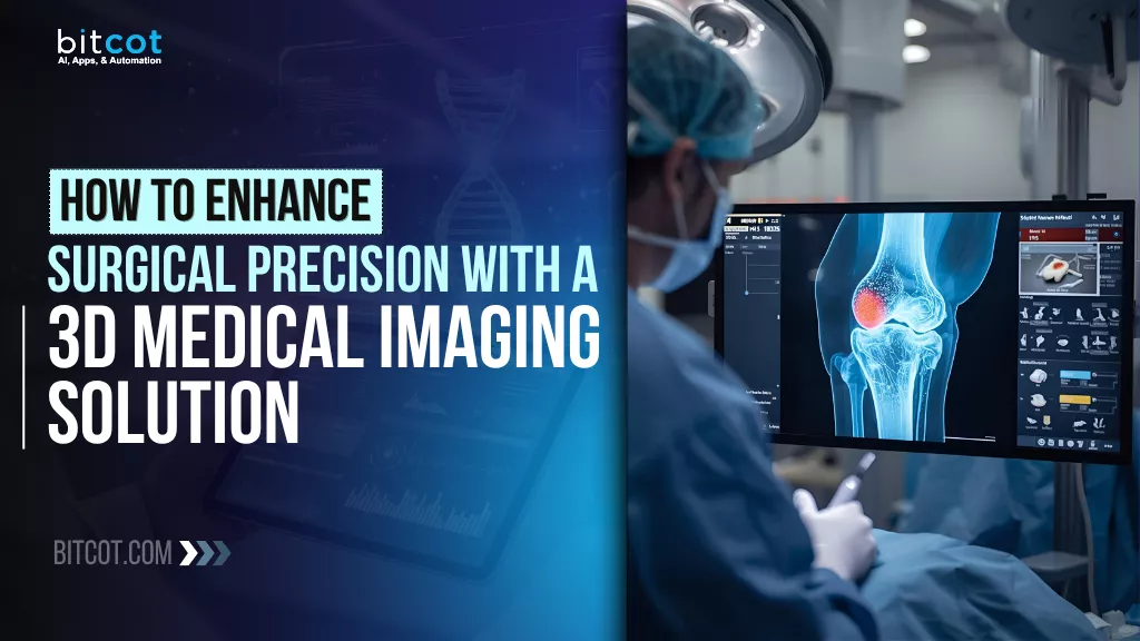
What is 3D Medical Imaging?
3D medical imaging is a modern approach to viewing and analyzing patient anatomy that moves far beyond the limitations of traditional 2D scans.
Instead of relying on flat images, where depth, angles, and spatial context must be mentally reconstructed, 3D medical imaging transforms standard DICOM data into interactive, lifelike 3D models that clinicians can explore in real time.
At its core, 3D medical imaging reconstructs cross-sectional slices from CT, MRI, or other imaging modalities into a volumetric representation. This means every structure, such as bone, tissue, or organ, can be examined from any angle, providing a level of clarity and accuracy that simply isn’t possible with conventional 2D views.
For clinicians, this unlocks significant advantages:
- True anatomical depth and realism: Instead of interpreting countless flat slices, they can visualize a patient’s anatomy exactly as it exists in three-dimensional space.
- Better understanding of complex structures: Overlapping tissues, intricate bone pathways, and challenging surgical zones become easier to interpret and navigate.
- More informed planning and decision-making: Surgeons can observe variations, anticipate challenges, and plan every move with a high level of precision.
- Enhanced collaboration across teams: 3D models create a shared visual language that reduces ambiguity and improves communication during case discussions.
The value of 3D imaging extends across almost every step of the clinical workflow. Radiologists gain more diagnostic accuracy. Surgeons gain more confidence in planning. Trainees gain clearer educational insights. And patients benefit from safer, more personalized treatment.
From a business perspective, 3D medical imaging represents a strategic leap forward. It reduces the cognitive load on clinicians, shortens planning time, lowers the risk of complications, and helps healthcare organizations deliver a higher standard of care.
As medical procedures become more complex and patient expectations continue to rise, 3D imaging is rapidly becoming a must-have technology, not just a nice-to-have upgrade.
In short, 3D medical imaging turns detailed medical data into clear, actionable insight. It bridges the gap between information and understanding, helping clinicians make smarter decisions with greater precision and confidence.
| Category | Traditional 2D Medical Imaging | Modern 3D Medical Imaging |
| Depth & Spatial Understanding | Limited depth; clinicians must mentally reconstruct anatomy | Full anatomical depth with accurate 3D visualization |
| Clarity of Complex Structures | Overlapping tissues make interpretation difficult | Complex structures are separated and clearly defined |
| Surgical Planning | Requires manual estimation and multiple cross-sections | Enables precise, interactive planning with realistic models |
| Implant Placement Accuracy | Higher chance of misalignment due to limited visibility | Enhanced precision with trajectory simulation and 3D mapping |
| Workflow Efficiency | Time-consuming; requires scrolling through many slices | Faster insights with one interactive 3D model |
| Error Reduction | Higher risk of misinterpretation | Lower risk due to complete spatial awareness |
| Team Collaboration | Difficult to convey complex anatomy | 3D models act as a shared visual understanding |
| Training & Education | Steeper learning curve for new clinicians | Intuitive and easier to learn and teach |
| Patient Communication | Harder for patients to understand 2D scans | Improves patient understanding and confidence with visual clarity |
Challenges of Traditional 2D Medical Imaging in Surgical Planning
Traditional 2D medical imaging has served the industry for decades, but its limitations become clear the moment surgeons begin planning complex procedures, especially those requiring high precision, such as orthopedic implant placement.
What appears to be a technical constraint on the surface quickly becomes an operational and clinical risk.
Medical professionals frequently report the same struggle: they are expected to plan highly intricate trajectories and placements while looking at flat images that offer no real depth. Without true 3D context, every decision becomes more difficult and time-consuming.
The core challenges are consistent across healthcare organizations:
- Significant limitations in complex procedures: When planning orthopedic implants or screw trajectories, 2D imaging simply cannot represent anatomical structures with the accuracy these procedures demand.
- Lack of depth perception and spatial clarity: Surgeons must infer depth, angles, and spatial relationships that are not visible in a 2D view, increasing the cognitive burden and the likelihood of error.
- No integrated surgical planning environment: With conventional tools, clinicians are forced to piece together information from multiple sources, making critical decisions with incomplete spatial data.
- Higher operational and clinical risk: These limitations often lead to suboptimal implant positioning, prolonged operative time, and a higher likelihood of complications during screw implantation.
For executives, these challenges impact far more than the surgical outcome; they affect efficiency, resource allocation, throughput, and overall quality of care. As procedure complexity increases, the gap between what clinicians need and what legacy imaging can provide becomes impossible to ignore.
This is exactly why organizations are exploring the shift toward advanced 3D medical imaging, and why we developed this POC to demonstrate what modern, integrated visualization can achieve.
How 3D Medical Imaging Transforms Clinical Practice
When clinicians have access to a complete, accurate, and interactive view of patient anatomy, the entire surgical planning experience changes.
Instead of working around the limitations of 2D imaging, they can operate with confidence, clarity, and precision, directly impacting outcomes and operational efficiency.
Our POC was built to demonstrate exactly how this transformation happens.
By integrating multi-view visualization, real-time 3D rendering, and intelligent trajectory planning into a single environment, the solution empowers surgical teams with tools that directly address the gaps traditional imaging leaves behind.
Here’s how it elevates clinical practice:
- Precise, data-driven surgical planning: Multi-view visualization gives clinicians synchronized perspectives of patient anatomy, allowing them to understand depth, angles, and spatial relationships with far greater accuracy.
- Intelligent implant trajectory planning: Instead of estimating screw paths or implant angles, surgeons can rely on automated guidance to visualize optimal trajectories before entering the operating room.
- Real-time 3D analysis: Surgeons can manipulate the 3D model instantly, rotating, zooming, and exploring anatomical structures, to refine decisions with the clarity that 2D imaging cannot provide.
- Automated screw recommendations: Built-in logic recommends screw length and placement based on the patient’s anatomy, reducing planning time and minimizing guesswork.
- Precise implant positioning controls: With the ability to fine-tune implant placement in a controlled digital environment, clinicians can ensure accuracy before making the first incision.
For business leaders, the impact is clear: better planning reduces surgical complications, accelerates workflows, and improves both patient outcomes and overall care quality. These improvements also support strategic goals, reducing operational risk, improving resource utilization, and strengthening the organization’s position as a forward-thinking healthcare provider.
This POC demonstrates how the right technological investment can dramatically elevate the standard of care, without requiring a full-scale system overhaul.
How 3D Medical Imaging Works in Surgical Planning
Behind the simplicity of an intuitive 3D visualization interface lies a highly sophisticated architectural foundation. For healthcare organizations evaluating advanced imaging capabilities, understanding how the technology works is key to recognizing its strategic value.
Our POC was engineered with a modern, scalable, and secure structure designed to handle complex medical datasets while delivering real-time performance.
Here’s a clear breakdown of how the system operates:
1. Architecture Powered by Cornerstone.js and Orthonac Integration
The system uses Cornerstone.js as the primary visualization engine, chosen for its ability to render medical images with high performance and reliability. To ensure secure and compliant handling of medical files, Cornerstone.js is integrated with Orthonac servers.
This combination allows the system to process, store, and manage DICOM data safely while supporting the advanced visualization features clinicians need.
2. Intelligent Data Processing Pipeline
The workflow begins as soon as a user uploads a DICOM file. The system automatically parses the study instance and identifies the relevant series within the file.
This structured pipeline eliminates manual sorting and helps clinicians access the exact datasets they need with minimal effort.
Once the data is processed, the rendering engine generates multiple viewports simultaneously. Users can view different orientations, such as axial, sagittal, coronal, and additional perspectives, at the same time.
Each viewport is synchronized, meaning any movement or adjustment in one view (such as zooming, panning, or scrolling through slices) is mirrored across all others for consistent spatial alignment.
4. Real-Time 3D Rendering and Surgical Planning Tools
Using advanced volume-rendering algorithms, the system transforms 2D slices into an interactive 3D model that can be manipulated in real time. Clinicians can rotate, explore, and analyze the anatomy without lag.
Within this same environment, the surgical planning module provides trajectory analysis and implant simulation. This allows users to map screw paths, test implant placements, and assess angles directly within the 3D model, ensuring accuracy before entering the operating room.
Benefits of 3D Medical Imaging in Surgical Planning
The shift from traditional 2D imaging to advanced 3D visualization delivers meaningful clinical and operational advantages.
By giving clinicians a clearer, more accurate understanding of patient anatomy, 3D medical imaging directly improves decision-making and enhances the overall quality of care.
At the core, the benefits center on one key outcome: greater precision.
1. More Accurate Diagnoses
With a fully interactive 3D view, clinicians can examine anatomical structures from every angle, reducing ambiguity and improving diagnostic confidence. Subtle variations that might be missed in 2D images become clear and measurable, helping teams identify issues earlier and with greater accuracy.
2. Tailored Treatment Plans for Each Patient
3D visualization allows medical professionals to build treatment plans that match the patient’s exact anatomy. Whether planning implant placement or navigating complex structures, the ability to personalize decisions leads to better outcomes and fewer complications.
3. Higher Precision in Surgical Planning
By providing a comprehensive spatial understanding, the system helps clinicians plan procedures with remarkable detail. Surgeons can map trajectories, simulate placements, and verify angles, resulting in more predictable results in the operating room.
4. Improved Patient Safety and Well-Being
Better planning leads to fewer intraoperative surprises and reduced risk. When implants are positioned accurately and procedures are executed with confidence, patients experience smoother recoveries, fewer complications, and overall better care.
For executives, these benefits translate into improved clinical performance, higher patient satisfaction scores, and a measurable reduction in avoidable surgical risks, strengthening both operational efficiency and patient trust.
Future Enhancements Planned for Our POC Solution
As we continue refining this 3D Medical Imaging POC, our roadmap focuses on expanding functionality, improving usability, and incorporating new capabilities that align with evolving industry needs.
These planned enhancements demonstrate how the solution can grow into an even more powerful medical device software tool for surgical planning and clinical decision support.
1. Expanded Surgical Type Support
To better illustrate the flexibility of the platform, we plan to introduce additional surgical types, beginning with Pedicle Screw Fixation.
This enhancement includes the ability to:
- Add new surgical types
- Edit existing types
- List available surgical workflows
- Delete or replace outdated configurations
These improvements will help teams explore how the platform can adapt to different specialties and procedure types, showcasing its scalability.
2. Real-Time Socket-Based Upload Tracking
We are introducing a socket implementation to track the DICOM upload process in real time.
This enhancement will:
- Improve visibility during file transfer
- Provide instant feedback to users
- Reduce wait-time uncertainty
- Enhance the overall user experience
For clinical teams dealing with large imaging datasets, this real-time tracking creates a more reliable and predictable workflow.
- New Cornerstone Toolkit Features
As Cornerstone releases new tools, we plan to integrate them to maintain alignment with industry advancements. Upcoming additions include:
- Planar Freehand ROI Tool for more flexible region-of-interest selection
- Annotation Eraser for quick adjustments
- Invert Tool to improve contrast visibility
- Label Tool for clearer marking and categorization
- Annotation Selection & Locking for more controlled editing
- Custom Orientation Marker to reinforce anatomical context
These enhancements deepen the platform’s precision and usability, giving clinicians more control during planning.
4. R&D on Updated Segmentation Capabilities
We are exploring upgraded segmentation methods to support more accurate anatomical separation and visualization. Improved segmentation will allow clinicians to isolate specific structures, analyze them independently, and build more detailed treatment plans.
Partner with Bitcot to Build Your Custom 3D Medical Imaging Solution
At Bitcot, we understand that every healthcare organization faces unique challenges, from improving clinical outcomes to streamlining workflows and optimizing resources. That’s why we don’t just build off-the-shelf products; we partner with you to create custom solutions tailored to your specific needs.
Our POC project is just the beginning. Through a collaborative approach, we can help you develop a scalable, secure, and highly effective imaging solution that aligns with your organization’s goals, workflow, and patient care standards.
- Tailored Solutions for Your Needs: We don’t believe in one-size-fits-all. Our team works closely with you to understand your challenges and deliver a solution that solves them, whether that’s integrating 3D medical imaging with existing systems, building new capabilities, or enhancing surgical planning workflows.
- Cutting-Edge Technology: We stay at the forefront of medical technology, ensuring your solution incorporates the latest advancements in imaging, real-time data processing, and user-friendly design.
- Security and Compliance: Healthcare data security is paramount. We ensure that all solutions comply with industry standards and regulations, providing peace of mind that your system is both functional and secure.
- Proven Expertise: With years of experience delivering custom software solutions, our team at Bitcot understands how to build robust, scalable applications that enhance both clinical and operational outcomes. We have the technical expertise and the healthcare insights to guide your project from concept to completion.
By partnering with Bitcot, you’re investing in more than just a tool; you’re investing in a long-term, flexible solution designed to grow with your organization’s evolving needs. Whether you’re looking to upgrade existing workflows or create entirely new ways to interact with patient data, our team is ready to help you bring your vision to life.
Let us help you transform the way your organization plans and executes surgeries, enhances patient care, and drives operational excellence. The future of 3D medical imaging is within reach, and Bitcot is here to make it happen.
Final Thoughts
3D medical imaging is more than a technical enhancement; it’s a smarter, clearer, and more confident way for clinicians to understand their patients. When surgical teams can see more, they can do more.
And that means better decisions, better outcomes, and ultimately, better care.
What this POC shows is simple: when you combine smart engineering with real clinical challenges, the result can be transformative. It becomes easier to plan with accuracy, easier to collaborate, and easier to deliver the kind of care modern healthcare demands.
The tool is designed to revolutionize diagnostics and treatment planning, empowering clinicians with deeper insights and supporting safer, more precise procedures. And because it’s built to evolve, it has the potential to drive ongoing medical innovation through continuous development.
And here’s the best part: solutions like this aren’t reserved for the biggest hospitals or the largest tech teams. With the right development partner, they’re completely within reach.
Whether you’re exploring new capabilities, evaluating healthcare digital transformation opportunities, or simply looking for a better way to support your clinicians, now is the perfect time to rethink what’s possible with the right technology.
At Bitcot, we help organizations turn complex ideas into practical, high-impact solutions. If you’re ready to explore what a tailored imaging solution could look like for your organization, we’d love to help you build it.
Partner with Bitcot for custom medical imaging software development services, and let’s create the future of healthcare imaging together.
Get in touch with our team.